The diagnosis and treatment of diseases is a delicate science. However, many people rely on home remedies to treat any discomfort or symptom that causes ill health. In this modern era of Google and search engines, it only takes a few seconds to find different alternative remedies to many conditions.
The thing about this type of approach is that many ends up treating illnesses they never had.
In such cases, any inaccurate assumption of an existing health problem means the actual disease has time to grow and eventually poses severe hazards to the patient’s health. This and other reasons are why it’s essential to carry out scientific and medically approved tests to diagnose a disease. One such test is the Elisa test.
The following sections will explain what the Elisa test means, its principles, and its methods.
What is Elisa?
Elisa is an abbreviated word that stands for enzyme-linked immunosorbent assay. It is essential mainly for research, food safety testing, and healthcare purposes. The Elisa enables the measurement of target analytes like antibodies, hormones, and protein biomarkers. Simply put, it’s used to detect the antibodies present in fluids like blood.
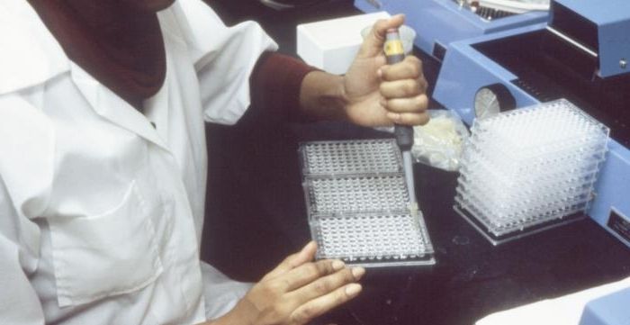
First described by Engvall and Perlmann in 1971, scientists also use it to study protein samples stationed in microplate wells by deploying particular antibodies. Typically, this is carried out in 384-well or 96-well polystyrene plates where the passive binding of proteins and antibodies occurs.
The process of immobilization and binding of reagents is why Elisa is simple to perform and design. This also gives rise to different types of ELISAs, such as the direct, indirect, and sandwich Elisa.
Sandwich ELISA
The sandwich Elisa enables the analysis of antigens between capture and detection antibody. The primary antigen under consideration must contain no less than two antigenic sites that readily bind to antibodies.
Usually, monoclonal or polyclonal antibodies can serve as the two layers between which the target antigen is measured in sandwich Elisa systems.
This Elisa eliminates the sample purification step before antigen analysis and boosts sensitivity about two to five times indirect or direct Elias. Other advantages include:
- Ideal for crude and complex samples
- Highly flexible
Here are five principles and methods of sandwich Elisa.
1. Coat With Capture Antibody
The procedure for the first sandwich Elisa principle is as follows:
- Use the capture antibody to coat the wells of a PVC microtiter plate at a concentration of 1–10 μg/mL in a buffer of carbonate/bicarbonate at pH 9.6.
- Then use an adhesive plastic to cover your plate and incubate at 4°C overnight.
- Next, take away the coating solution and fill the wells with 200 μL PBS to clean the place twice. The washing solutions are rinsed off after flicking the plate over a sink. The drops left on the plate are removed with a paper towel by dabbing.
2. Adding Samples And Blocking Them
- Add 200 μL blocking buffer (5% non-fat dry milk/PBS) to each well to block the rest of the protein-binding sites of the coated wells.
- With an adhesive plastic, cover the plate for about one to two hours at room temperature or a temperature of 4°C overnight.
- Thoroughly rinse the plate with 200 µL PBS at least twice.
- Add to each well 100 μL of diluted samples. Always measure the differences between the signal of unknown samples and the signals of a standard curve. And run duplicates or triplicates and a blank on every plate, then incubate at 37°C for 90 minutes.
- Take out the samples and bathe each plate in 200 μL PBS twice.
3. Adding Detection And Secondary Antibodies For Incubation
- Add to each well a diluted detection antibody (100 μL). Verify that the detection antibody perceives a separate epitope on the target protein to the capture antibody.
- Use an adhesive plastic to cover the plate and incubate at room temperature for two hours.
- Next, use PBS to bathe the plate four times.
- Get and dilute 100 μL of conjugated secondary antibodies in the blocking buffer and immediately add it to your setup.
- Return the adhesive cover of your plate and incubate at room temperature for one to two hours.
- Again, use PBS to wash the plate four times.
4. Identification
Alkaline phosphatase (ALP) and horseradish peroxidase (HRP) are two of the most commonly used detection enzymes in Elisa assays.
- ALP Substrate
Most applications use P-Nitrophenyl-phosphate (pNPP). Record nitrophenol’s yellow color at 405 nm after 15 to 30 minutes incubation at room temperature.
Then add an equal amount of 0.75 M NaOH to end the reaction.
- HRP Chromogenic
Hydrogen peroxide is the substrate of HRP. Its cleavage is combined with the oxidation of a hydrogen donor resulting in a change in color during the reaction.
- TMB
Add to each wall some TMB solution, incubate for 15-30 min, add the same amount of stopping solution and record the optical density at 450 nm.
- OPD
The measurement for the final product is 492 nm. The substrate is light-sensitive so store it in the dark.
- ABTS
The final product has a green color, and the reading for its optical density is 416 nm. Remember never to handle carelessly and always wear gloves because some enzyme substrates are carcinogens and could be hazardous.
5. Data Analysis
Produce a standard curve using the serial dilutions data by placing concentration on the x-axis and absorbance on the Y-axis. Using this standard curve, interpolate the sample’s concentration.
Conclusion
Apart from this application and the uses of Elisa that were covered in this post, Elisa technology can be found in standard diagnostic kits. Some of these kits you can buy over the counter. A good example is the home pregnancy test kit. The general term for these tests is “dip-stick” Elisas. They also apply the sandwich Elisa principles and use lateral flow. This is but one of the many instances that Elisa provides rapid and reliable test results. However, the results from these simplified Elisa types lack the quantifiable quality.